





Low - Moderate

LUMBAR DISC HERNIATION

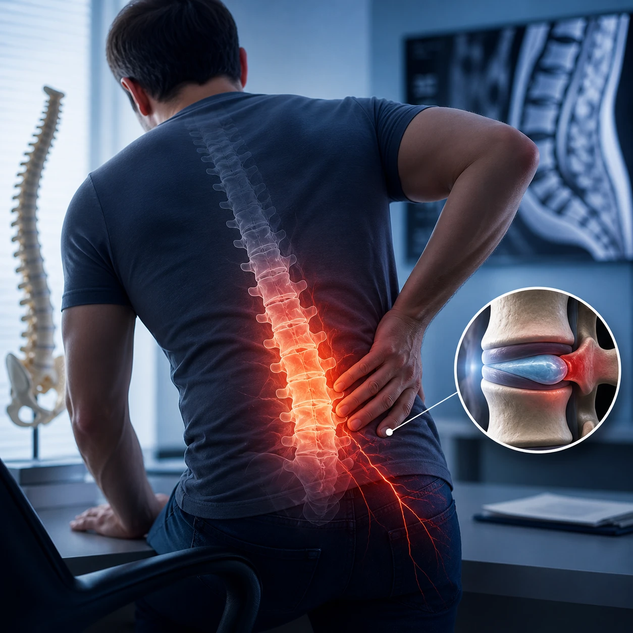
Lumbar disc herniation is a condition that occurs when the discs in the lower back region of the spine shift out of place or tear.

What Is Lumbar Disc Herniation?

Lumbar disc herniation is a condition that occurs when the discs in the lower back region of the spine shift out of place or tear. The discs located between the vertebrae in the spine help facilitate movement and provide protection against impacts. When the tough outer layer of these discs is damaged or weakened, the soft inner part may protrude outward and put pressure on the nerves.

What Causes Lumbar Disc Herniation?

Lumbar disc herniation may generally occur as a result of excessive strain, heavy lifting, or sudden movements. In addition, sitting or working in poor posture for long periods may also cause lumbar disc herniation. During the aging process, the loss of elasticity and durability of the spinal discs increases the risk of lumbar disc herniation. Excess weight may also trigger this condition by placing extra pressure on the spine. Genetic predisposition is also an important factor that may lead to lumbar disc herniation. In other words, individuals with a family history of lumbar disc herniation have a higher risk of developing this condition.

What Are the Symptoms of Lumbar Disc Herniation?

Lumbar disc herniation may present with pain in the lower back area. This pain may become more severe, especially during sudden movements or when remaining in the same position for a long time. If it puts pressure on the nerves, the pain may start from the hip and spread down to the legs, known as sciatic pain. In addition, numbness, tingling, or weakness may occur in the legs and feet. In some cases, limited mobility may be experienced in the affected area, making daily activities difficult. In severe cases, difficulty walking or balance problems may also occur.

How Is Lumbar Disc Herniation Treated?

  • Medication Treatment: Painkillers, muscle relaxants, and anti-inflammatory medications.
  • Physical Therapy: Exercises to support the spine and reduce pressure on the nerve.
  • Non-Surgical Treatments: Thanks to modern medical methods such as ozone therapy, radiofrequency, or laser discectomy, the herniated disc is accessed with a thin needle and reduced in size. “Non-Surgical Lumbar Disc Herniation Procedures”
  • Surgical Intervention: Surgery to relieve pressure on the disc in severe cases.

Surgical Treatment of Lumbar Disc Herniation

Discectomy is the most commonly used surgical treatment for lumbar disc herniation. Intervertebral discs are structures located between the bones of the spine, connecting the vertebrae and firmly attached to them. When the outer surface of the disc, called the “annulus fibrosus,” weakens due to age and injury, tears may occur. The soft inner part of the disc, which provides flexibility and is called the “nucleus pulposus,” may deteriorate, shift out of place, and protrude outward through tears in the outer layer. The slipping, sagging, or bulging of the disc is called herniation. In medical literature, disc herniation is referred to as a herniated disc. When the inner disc material extends beyond the posterior outer surface boundary, it may put pressure on very sensitive nerve tissue. The protruding disc may compress or damage the nerve root, resulting in pain radiating to one or both legs, as well as loss of strength and numbness depending on the affected nerve root or roots. Discectomy is performed to remove herniated disc fragments and damaged parts, thereby relieving pressure on the nerve root and reducing pain.

Disc surgery involves a small incision in the skin over the upper part of the spine, removal of some ligament and bone material, and removal of disc fragments.

Open discectomy has been performed for the last 60 years. Physicians have improved this surgical approach by making better surgical plans with advanced diagnostic methods such as magnetic resonance imaging (MRI) and computed tomography (CT).

In microdiscectomy, a smaller skin incision of approximately 1.5–2 cm is made. Muscle tissue is minimally separated, and less bone is removed.

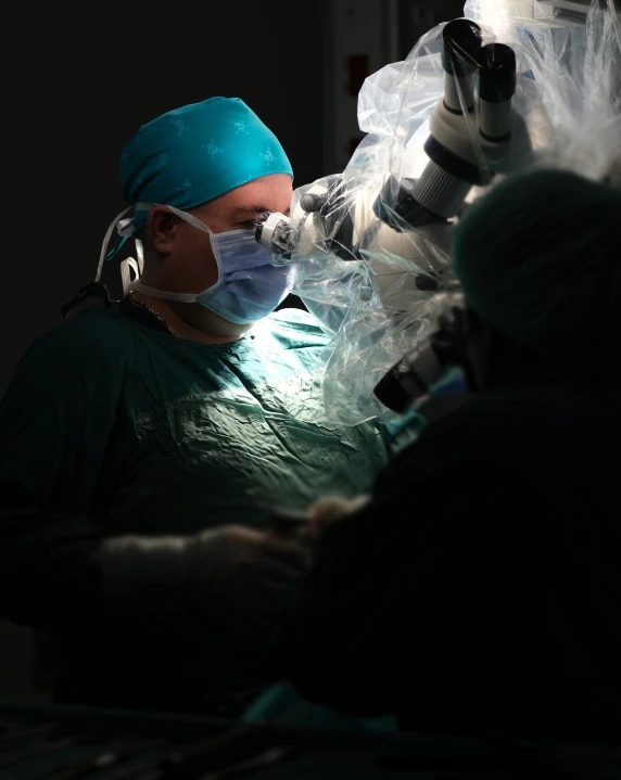
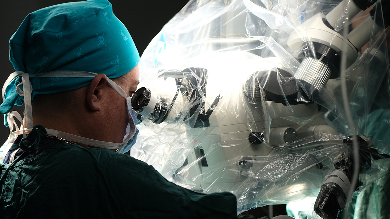
Since microdiscectomy is performed using a microscope and microsurgical instruments, the risk of nerve injury is quite low. After surgery, the patient can return to work more quickly.

WHO NEEDS DISCECTOMY?

Not all patients with disc herniation are candidates for open discectomy. In most people, pain relief can be achieved with conservative treatments such as rest, physical therapy, analgesic and anti-inflammatory medication use, and epidural injections. However, sometimes pain does not respond to these treatments, and surgical interventions may be required.

If lower back or leg pain does not respond to conservative treatments and continues for 4–6 weeks or longer, physicians may request diagnostic tests and imaging methods such as X-ray, MRI, or CT to identify the source of the pain. If the diagnosis of disc herniation is confirmed, open discectomy or microdiscectomy may be recommended.

Some disc herniations can be removed endoscopically through a smaller incision using special instruments under local anesthesia. However, open discectomy or microdiscectomy is still considered the “gold standard” for the surgical treatment of disc herniation, especially when disc protrusion becomes prominent or when a disc fragment breaks off and causes significant pain and weakness. Open discectomy allows the surgeon to see and explore the surgical area in the best possible way.

HOW ARE OPEN DISCECTOMY AND MICRODISCECTOMY PERFORMED?

Open discectomy is usually performed under general anesthesia, with the patient asleep and unconscious, and typically requires a one-day hospital stay. It is performed while the patient is lying face down or in a knee-elbow supported crouched position. The procedure is carried out through an incision of approximately 2–4 cm made in the skin over the affected area of the spine. Muscle tissue is separated from the bones above and below the affected disc, and a retractor moves the muscle and skin away from the surgical area so that the surgeon can see the spine and disc more clearly. In some cases, bone and ligaments may be removed to provide a better view, allowing access to the bulging and damaged disc without injuring the nerve root. This procedure is called laminectomy or laminotomy, depending on the amount of bone removed. In this way, the surgeon can see the vertebra, disc, and surrounding structures. After these steps, the disc fragments protruding from the disc wall and removable disc fragments are removed.

In microdiscectomy, this procedure is often performed under magnification using a surgical microscope. A smaller incision is made, muscle tissue is separated less, and a better view is obtained.

No material is used to replace the removed disc tissue. The incision is then closed with sutures, and the patient is taken to the recovery room.

WHAT HAPPENS AFTER SURGERY?

After surgery, you may feel pain in the incision area, and the pain may not completely disappear immediately after the operation. Your doctor may arrange pain management to make your postoperative process more comfortable. You will learn deep breathing techniques and may be asked to cough in order to clear secretions from your lungs that may occur due to general anesthesia. It is recommended that you start walking as soon as you recover from anesthesia, usually after 6 hours, and have a companion to help with your recovery.

Before leaving the hospital, you will be informed about what to pay attention to in order to feel comfortable during activities such as climbing stairs, sitting, and getting in or out of a car or bed. When you are discharged from the hospital, your doctor may arrange physical therapy suitable for your condition.

During the first 4 weeks after surgery, certain restrictions may be recommended, such as avoiding prolonged sitting, not lifting objects heavier than 5 kilograms, and avoiding excessive bending or stretching. You should also not attempt to drive until your doctor gives permission.

WHAT ACTIVITIES CAN I DO AFTER SURGERY?

Walking is the first physical activity you will try. Walking reduces the risk of tissue scarring in the surgical area and helps maintain spinal mobility. Within a few weeks, you may also be allowed to cycle and swim. Proper physical therapy can maximize your recovery.

People working in jobs that do not require physical effort may return to work within 2–4 weeks or even sooner. Those working in jobs involving heavy lifting or operating strong vibrating machinery may need to wait 6–8 weeks after surgery before returning. Physical therapy may play a role in your recovery.

ARE COMPLICATIONS POSSIBLE?

Possible complications of open discectomy include bleeding, infection, cerebrospinal fluid leakage, injury to arteries and veins near the spine, and damage to the nerve root or the surrounding protective layer called the dura mater.

In addition, recurrent disc herniation may occur in the same disc after surgery. Recurrent disc herniation is seen in approximately 5–10% of cases after open discectomy.

All surgical methods carry a risk of complications, and all risks should be discussed with your surgeon before the surgical procedure. Excessive bleeding, redness, discharge from the surgical site, fever, weakness, numbness in the legs, or urinary problems are certain warning signs. If you experience any concerning symptoms, inform your doctor.

ARE COMPLICATIONS POSSIBLE?

Medical studies have shown an 80–90% success rate in cases requiring open discectomy treatment. This rate is slightly higher in microdiscectomy. According to studies, patients with radicular pain, which is pain radiating to the legs, benefit more than those who undergo open discectomy due to lower back pain complaints.

To increase effectiveness, open discectomy is suitable for people experiencing persistent, severe hip and leg pain or weakness. If your lower back or leg pain is not at this level, open discectomy may not be a recommended method for you; in that case, you may improve with conservative treatments or less invasive therapies.

If you have lower back or leg pain, before any method is applied, you should discuss your pain with your doctor and evaluate all possible treatment options.

```

symptoms

  • Lower back pain
  • Leg pain and numbness
  • Loss of strength in the leg
  • Difficulty sitting for long periods

Risk Factors

Heavy lifting Sedentary lifestyle Smoking Genetics

Make a Quick Appointment

Early diagnosis saves lives. Fill out the form to plan your treatment.

Similar Diseases

Cookie Policy As Op. Dr. Selçuk Gözcü, we use cookies on our website. This Cookie Policy (“Policy”) applies to the website https://selcukgozcu.com/, managed by Op. Dr. Selçuk Gözcü, and cookies will be used as specified in this Policy.

I Accept